 Character identification
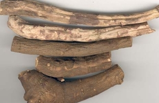
 The dried roots are cylindrical, with both ends roughly equal in thickness, slightly curved, 10 to 36 cm long, and about 6 to 19 mm in diameter. Dark brown or dark brown surface, rough, with lateral convex lenticels and root marks, with thick and deep vertical wrinkles, hand peeling the skin is easy to fall off, showing a white or light brown skin. Hard and brittle, easy to break. The section is flat, pinkish-white or yellowish-white, the cortex is narrow, pinkish-like, the central medullary part is small, the ray of the essential part is obvious, and sometimes it has fissures. Gas slightly fragrant, slightly bitter taste. Thick and long roots, skin easily fall off, thick and deep wrinkles, cross-section white, pink is better. It is mainly produced in Inner Mongolia, Hebei, Liaoning, Heilongjiang, Jilin, Shaanxi, Shanxi, Gansu, Qinghai, Hubei, Sichuan, Guizhou and Yunnan. Among them, red peony roots produced in the northeast, north China and northwest regions are mainly roots of peony roots, and some are roots of grass peony roots. The red peony root medicine produced in the southwest area is dominated by the roots of the Sichuan red peony root.
Among the above products, the quality produced in Duolun, Inner Mongolia is the best, and it is known as “Duolun Akasakaâ€. Producers from Shaanxi and Gansu still have the roots of the paeonia anomala l., which is mostly in the form of a spindle. In Sichuan, in addition to Chuan Chia, some regions also use the roots of the beautiful p.mairei levl. and p.obovata maxim.var.willmottiae(stapf)stern. Sichuan products are divided into two types, shaved red peony and raw red peony. The shaved skin red peony is removed from the skin and is cylindrical. The surface is pale reddish purple or flesh-white, with forward wrinkles. The outside of the section is lavender, and the inside is light yellow. Rays. The original skin red peony is not peeled, similar in shape, but there are fork branches, the appearance of rough brown red or brown. Producers from Xinjiang, in addition to the above-mentioned grass peony, also use p.hybrida pall., whose roots are spindle-shaped.
Microscopic identification
(1) Paeonia lactiflora: Root cross-section: Cork layer is 5-10 cork cells; there is residual skin layer. The cortex is narrow, and some cells have separate mother and daughter cells. The phloem sieve group is evident at the near formation layer, and some phloem inside the tube. The formation of a microwave-like ring. Xylem rays are broad; single or clusters of conduits are arranged in the same order as wood fiber bundles; central conduits are grouped with wood fibres in two groups. Large pits are visible in the cortex, phloem, and ray parenchyma cells. The product parenchyma cells contain starch grains, and some contain calcium oxalate clusters.
Powder: light brown red.
1 Calcium oxalate clusters are often arranged in a vertical row, with a diameter of 7-38 (-41) μm, with small crystal-containing cells and curved walls. Some cells contain two or more crystals.
2 Fibrous tracheid fusiform, diameter 14-38μm, wall thickness 5-13μm, bordered pits larger, pitted mouth slit-shaped, there are wide pits and cross into a cross-shaped; a few tough type Fiber with a single twill hole.
3 The cork cells have a long, rectangular, or long polygonal surface, with a length of approximately 225 μm; some cells are filled with brown or reddish-brown lumps.
4 The bordered pits are 25-78μm in diameter, with marginal ellipsoids, and some extend laterally to form mesh or ladder-shaped bordered pits. Perforation plates are located on the end wall or side wall, with 1-4 perforations. Another starch grain, diameter of about 15μm.
(2) Chuan Chika: Root cross section: The skin layer is sometimes visible. The cortex is narrow. The phloem sieve group is not obvious. Form a layer of ring wave. There are more xylem vessels near the formation layer, scattered in a few or a few groups; wood fibers and catheters between the birth; a small number of central catheter and wood fibers scattered. Cortical and phloem can sometimes be seen in tubular closed tissue with central parenchyma cells containing brown-red secretions. The product parenchyma cells contain starch grains, and some contain calcium oxalate clusters.
Powder: brown.
1 The tubular closed tissue fragments show that the central parenchyma cells contain brown-red material.
2 fiber tracheid diameter 15-30μm; tough fiber diameter 14-36μm.
3 The starch diameter is approximately 21 μm.
Chemical identification
(1) Cross section of the product: The cork layer is a series of brown cells. Cortical parenchymal cells are tangentially elongated. The phloem is narrow. Layer formation loop. The xylem rays are wide, the catheter groups are arranged radially, and there are wood fibers next to the catheter. Parenchyma cells contain calcium oxalate clusters and contain starch grains.
(2) Take the product powder 0.5g, add ethanol 10ml, shake for 5 minutes, filter, the filtrate evaporated, the residue plus 2ml of ethanol to dissolve, as the test solution. Separately, the reference substance of paeoniflorin was taken, and ethanol was added to make a solution containing 2 mg per 1 ml as a reference solution. According to the thin layer chromatography (Appendix VIB) test, 4 μl of each of the above two solutions were pipetted onto the same silica gel G plate, using chloroform-ethyl acetate-methanol-formic acid (40:5:10:0.2) as the The developing agent is developed, taken out, dried, sprayed with a 5% vanillin sulfuric acid solution, and heated until the spots are clearly colored. In the chromatogram of the test sample, the same blue-purple spot was observed at the position corresponding to the chromatogram of the reference substance.
Frozen Squid Products,Frozen Squid,Frozen Calamari Rings,Frozen Squid Woolworths
Zhoushan Fudan Tourism CO., LTD , https://www.fudanfood.com